Saturday, July 25, 2009
Lab Technique - Electrochemistry
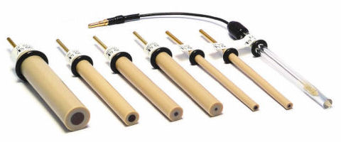
Electrodes come in different sizes and differ in the inert material in the middle. This picture shows a few glassy carbon electrodes. For a gold electrode, the middle will be gold instead of grey.
Electrodes are electrical conductors that can be used for various purposes such as electrocardiography (ECG) and chemical analysis using electrochemical methods. There are different types of electrodes available in the market but the ones that I have been using are the gold (Au) electrodes.
In order to ensure that the Au electrodes are fit for use, they are polished to remove unwanted chemical residues from previous use. New electrodes of course, do not need to be polished. Polishing is done using alumina powder on a nylon pad. The electrodes are held perpendicular to the nylon pad (with Au in contact with the pad that has alumina powder.) Then for a desired time (eg 10 min each), these electrodes are polished in a figure of 8 or circular motion. The particles in the alumina powder will remove the residues on the gold surface. In addition, nano-strip can be used too. By incubating the electrodes in nano-strip for 15 min, any remaining organic residues are removed.
So how do you know if the electrodes are clean? Electrochemical signals are measured with an electrochemical workstation. A clean Au electrode should give a high electrochemical signal while a Au electrode covered with residues will give a low signal because these residues will slow down the transfer of electrons between the electrode and electrolyte.
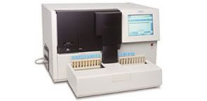
This is a picture of an electrochemical workstation. The electrodes are connected to the workstation with the use of wires so that a small amount of voltage can be passed through.
Yvonee Chew 0703189A
Sunday, July 19, 2009
Cytology - Processing Gynaecological Specimens
it's already end of week 4 of SIP. 16 weeks more to go. 4 mths more to go!
anyway. i'm attached to the histopathology/cytology department. over at the hospital i'm attached to, histopathlogy actually sort of belong to the same department, though both deals with different type of specimens, but is usually inter-related.
we didn't get to do much during our first week, even though for that whole week we were at histo department. from week 2 onwards, we are all allocated at different sections. as for me, i'm now at cytology lab for 1mth, so for this post, i'll be talking about things that are done at our cytology lab. =)
Cytology is actually the study of cells to aid in the diagnosis of diseases. In our lab, we deal with 2 different groups of specimen, Gynaecological (gynae) and Non-gynaecological (NG) specimens. Gynae specimens are actually just cervical pap smears, while NG specimens refers to body fluids (peritoneal, pleural, pericardial, esophageal washings, bronchial washings/aspirates etc), CSF, sputum, urine, and fine needle aspirations (knee, breast, thyroid, bones etc). I'm now covering both gynae section and NG section, but due to safety reasons, we're actually not allowed to perform hands-on for NG specimens, so yeah, all we can do is observe and learn the techniques. =) I'll cover on the gynae section because that's what i've been doing most of the time.
Gynae specimens usually comes in two different type. Liquid-Based preparation or Conventional Smears.
Ang Yu Hui
0702632A
Thursday, July 9, 2009
Hb Electrophoresis
One of the routine work I had to do was sample processing. These are the few steps I had to follow:
1. Collect samples from the Haem reception counter.
2. Label request form and sample lab no, ensure patients particulars match with sample, initial.
3. Record patient's data (as well as all results) in Hb Electro worksheet.
4. Set up cellulose acetate electrophoresis.
5. Set up HbH inclusion bodies test.
6. Record patient's FBC from LIS
7. If no result in LIS, run FBC using the analyzer and file results.
Other than sample processing, I had to perform this test called Cellulose Acetate Electrophoresis. This test helps to detect common Hb variants present in the blood, if present. Cellulose Acetate is used as it is easily available, provides sharp resolution of Hb bands in a short time, and permits clearing.
Materials needed:
1. Supre-Heme buffer
2. Helena Hemolysate reagent
3. Ponceau S stain
4. 5% glacial acetic acid
Procedures:
1. Add 200ul of patients' sample into 10x75mm test tubes
2. Wash the cells by adding 0.9% saline till 3/4 full
3. Centrifuge at 3000rpm for 10 min
4. Repeat steps 3 & 4 to obtain packed cells
5. Dispense 25ul of hemolysate reagent in another set of 10x75mm test tubes
6. Add 25ul of packed cells into each of the test tubes containing hemolysate reagent (Note: Pipette up and down to ensure complete haemolysis)
7. Place 10ul of packed cells containing hemolysate reagent into each sample well of prime applicator
8. Prime applicator by depressing the tips gently into samples wells and blot on filter paper
9. Remove wet titan cellulose acetate plate from buffer and blot once between 2 blotters
10. Prime applicator again and transfer to the wet titan cellose acetate plate
11. Transfer the plate to the electrophoresis chamber with the acetate faced down
12. Electrophoresis the plate for 25 min at 350 volts
13. Staining of Hb bands occurs after 25 mins by placing the plates into Ponceau S stain for 5 min
14. Destain in 3 successive washes of 5% acetic acid (2 min for each wash)
On the first two weeks, I've been using the SYSMEX automated blood coagulation analyzer CA-1500.

Picture from http://diagnostics.siemens.com/webapp/wcs/stores/servlet/ProductDisplay~q_catalogId~e_-111~a_catTree~e_100001,1015818~a_langId~e_-111~a_productId~e_182051~a_storeId~e_10001.htm
I feel that this is a great invention! It can do multiple tests such as PT, APTT, D-Dimer Assay, Factor Assays, Protein C Assay, Protein S assay etc. The turnaround time for the tests ordered is fast and it increases productivity rate! However in the Stat Lab, only PT and APTT tests are done. The rest of the tests are sent to the Special Coagulation Lab to do.
So basically when samples arrive, this is what I'll do!
- Collect samples from the Reception Counter
- Remove blood tube from biohazard bag
- Check that the name tally with the request form
- Ensure correct anticoagulant tube is used (citrated ; light blue)
- Check that volume is sufficient
- Assign a lab serial number on the request form and initial name next to it
- Paste the respective barcode label with correct serial number and date on the blood tube
- Before spinning, invert tube to look for big clot
- Centrifuge @ 3000rpm for 3minutes. At the same time, log in request in LIS
- After centrifuging, place them on the sample rack and put on machine's right rack pool ensuring that the barcode labels are facing the scanner in the machine when running.
- Press START on the analyzer screen to run the test
- Record the results on the request form
- Update results in LIS
Therefore the principle of Prothrombin Time (PT) is to screen for abnormalities of factors involved in extrinsic pathway such as factor II, V, VII, X, prothrombin and fibrinogen whereas APTT is a screening test that measures clotting factor of intrinsic pathway such as VIII, IX, XI and XII. These can be easily done by using this automated analyzer to perform the tests! The most commonly ordered tests are PT and APTT. It makes work easier for us because manual PT/APTT is so time-consuming! Remember we did it during the Haem practical? Gosh, it's going to take us hours to complete all the tests ordered! Our lab only do it manually if the first result is out of range and when repeated, result is within the range.
Reference range for PT : 9.0 - 12.5sec
Reference range for APTT : 26.0 - 36.1 sec
The manual test is to confirm the result of the repeated test before recording it in the LIS. However, manual testing is rarely done in our lab!
And thats all for my first few weeks in the Stat Lab! I'll be in the Special Coagulation Lab this week and I'll be learning more other tests!
Till then!
-QINGLING- :D
Sunday, July 5, 2009
Immunoprecipitation (IP)
Materials
- Protein A/G plus Agarose – protein A/G are used with rabbit/mouse antibodies respectively
- 20X Coupling buffer
- Disuccinimidyl suberate (DSS)
- IP Lysis/Wash buffer
- 100X Conditioning Buffer
- 20X Tris-Buffered Saline
- Elution Buffer
- Non-reducing 5X Lane Marker Sample Buffer
- Spin Columns
- Microcentrifuge Collection Tubes
- Microcentrifuge Sample Tubes
- Control Agarose Resin – used to pre-clear lysates to reduce non-specific protein binding
- Ultrapure water
Note:
- Flow through rate should not exceed 600/300ml when using a 2/1.5ml collection tube respectively; as exceeding these volume may result in back pressure in the spin column/incomplete wash/incomplete elution.
- All resin centrifugation steps should be performed at low speeds i.e. 1000-3000 x g as high-speed centrifugation i.e. > 5000 x g may cause resin to clump and make resuspension difficult.
Method
(i) Binding of antibody to protein A/G plus Agarose
- Prepare 2ml of 1X coupling buffer for each IP reaction by dilution 20X coupling buffer with ultrapure water
- Using a cut pipette tip, add 20ml of resin slurry into a spin column and place the column into a microcentrifuge tube and centrifuge at 1000 x g for 1 minute. Then discard the flow through. Note: Gently swirl the bottle of pierce protein A/G plus agarose to obtain an even suspension
- Wash the resin twice with 200ml of 1X coupling buffer, centrifuge and discard the flow-through.
- Gently tap the bottom of the column on a paper towel to remove excess liquid and insert the bottom plug.
- Prepare 10mg of antibody for coupling by adjusting the volume to 100ml with sufficient volume of ultrapure water and 20X coupling bluffer. Note: Add the ultrapure water, 20X coupling buffer and affinity-purified antibody directly to the resin column.
- Attach the screw cap to the column and incubate on the rotator at room temperature for 30-60minutes; ensuring that the slurry remains suspended during incubation.
- Remove and retain the bottom plug and remove the cap. Then place the column in a collection tube and centrifuge. Note: Save the flow-through to verify antibody coupling.
- Wash the resin with 100ml of 1X coupling buffer, centrifuge and discard the flow-through.
- Wash the resin twice with 300ml of 1X coupling buffer, centrifuge and discard the flow-through.
Note: DSS crosslinker is moisture sensitive and incompatible with amine-containing buffers e.g. Tris, glycine. DSS should also be dissolved in DMSO/DMF immediately before use (dilute DSS solution in 1:10 DMSO/DMF to make 2.5mM)
- Tap the bottom of the column on a paper towel to remove excess liquid and insert the bottom plug.
- Add 2.5ml of 20X coupling buffer, 9ml of 2.5mM DSS and 38.5ml of ultrapure water to the column to make up 50ml. Then attach the screw cap.
- Incubate the crosslinking reaction or 30-60 minutes at room temperature on a rotator.
- Remove and retain the bottom plug and open the bottom plug and screw cap and place the coloumn into a collection tube and centrifuge.
- Add 50ml of elution buffer to the column and centrifuge. Note: Save the flow-through to verify antibody crosslinking.
- Wash four times with 200ml of elution buffer to remove non-crosslinked antibody and quench the crosslinking reaction.
- Wash twice with 200ml of cold IP lysis/wash buffer and centrifuge after each wash. Note: Antibody-crosslinked resin can be stored up to 5 days in IP lysis/wash buffer. For longer storage, store resin in 1X coupling buffer (PBS).
Note: If antibody-crosslinked resin was stored in PBS, wash twice with IP lysis/wash buffer and discard the flow-through after each wash.
- Tap the bottom of the column on a paper towel to remove excess liquid and replace the bottom plug.
- Add sample to the antibody-crosslinked resin in the column. Then attach the screw cap and incubate the column overnight at 4oC on the rotator.
- Remove the bottom plug, loosen the screw cap and place in the column in a collection tube and centrifuge the column. Save the flow through. Note: Do not discard the flow-through until confirming that immunoprecipitatio is successful.
- Remove the screw cap, and place the column into a new tube. Add 200ml of 1X TBS buffer and centrifuge.
- Wash sample twice with 200ml IP lysis/wash buffer and centrifuge after each wash.
- Wash sample once with 100ml of 1X conditioning buffer.
- Add 20ml of elution buffer into the column and incubate for 5minutes at room temperature. Centrifuge.

