Saturday, November 7, 2009

Hello everyone! Qingling here! SIP's over! :D
This is gonna be my last entry!


Alright, I was in the bone marrow lab for my last section.
but however, i will not mention about the bone marrow aspiration
as vanessa had done so in her blog post (group 6).
pssst! its my most enjoyable lab experience ever! :D


In the bone marrow lab, not only do we go down to the ward
for bone marrow aspiration, we also do staining of the slides and viewing and
reporting of the results. So in this entry, i'll describe to you guys the different
stainings!


Maygrunwald-Giemsa stain:
it is a romanowsky stain used in morphologic classification
of hematopoietic cells. Giemsa alone can demonstrate malarial parasites in thick film.
Romanowsky stain consists of methylene blue and oxidation product and eosin Y or eosin B.
The combined action of these dyes will produce the Romanowsky effect yielding a purple colour
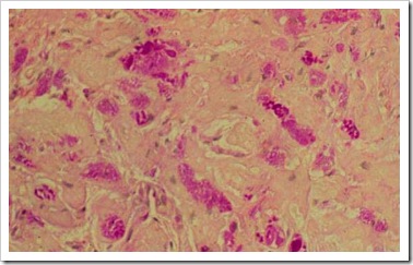
to nuclei of leukocytes and neutrophil granules, and pink colour to erythrocytes. M/G stained wedged bone marrow smear tells us the cellularity, quantity, quality of the magekaryocytes. Presence of tumour clumps should be noted when viewing slides. It also stains the erythroid and granulocytic cells, lymphoid, reticulum and plasma cells.

Procedure :
1) Add 1:1 ratio of Maygrunwald stain to buffered water onto the slides.
2) Mix well and leave it for 7 minutes.
3) Wash throughly.
4) Add 1ml of Giema stain adn 9ml of buffered water.
5) Mix well and leave it for 12 minutes.
6) Wash and air dry.

picture taken from http://www.bccancer.bc.ca/NR/rdonlyres/5A3C3A86-4549-4422-8258-BE8AE2635461/3748/adenogiantcell03208501w2.jpg


Iron stain :
The HCl caused ferric iron in hemosiderin to release from protein. The free iron react with potassium ferrocyanide and gives an insoluble blue compound called ferric ferrocyanide. The ferritin which is soluble does not give a positive reaction. An increase in iron stores shows more hemosiderin granules that are often in clumps on the slides. Hemosiderin is stained blue-green in colour while nuclei is stained red.


Iron stain procedure :
1) A control is used together with the first iron stain slide of the day.
2) Fix the slides with methanol for 10-20 minutes.
3) Wash it with water
4) Mix 1ml of HCL and 1ml of potassium ferrocyanide into a testtube and mix.
5) Pour the mixture onto the slides and leave it for 15 minutes.
6) Counterstain with neutral red solution for one minute.
7) Wash and air-dry


Picture taken from http://aphlabs.com/yahoo_site_admin/assets/images/DSCN8716.187192835_std.JPG


Periodic Acid Schiff stain :
It stains a variety of carbohydrates including glycogen which is often found in haemopoietic cells. It is used to differentiate the diagnosis of acute leukemia. Lymphoblast in coarse granules or large block on clear background are PAS positive. Myeloblast and monoblast are PAS negative. The product will be red in colour. Neutrophils are magenta and cytoplasm are red in colour. Normal erythroblasts do not stain PAS. However PAS is strongly positive in erythroleukemia.


Procedure :
1) Fix the slides with methanol for 15-20 minutes.
2) Wash and add periodic acid and stain it for 10minutes.
3) Rinse and AIR-DRY!
4) Add schiff reagent and leave it for 20 minutes
5) Rinse with water
6) Counterstain with Harris Haematoxylin for 10minutes.
7) Run in tap water for 5minutes.


Picture taken from http://www.kidneypathology.com/Im%E1genes/GNM/05-7007.PAS.2.w.jpg


Peroxidase Stain:
It establish and confirm the diagnosis of acute myeloid leukaemia since lymphoblasts are negative. It requires >3% of bone marrow blasts to show peroxidase activity for acute leukemia to classify as myeloid. Myeloloperoxidase deficiency produces clinical picture similar to that of chronic granulocytic disease. Active myeloperoxidase in granulocytes implies that opsonin function is intact. Cytoplasm shows golden yellow granules with nuclei purple in colour. Monocytes, mature and immature granulocytes and many myeloblasts are positive for peroxidase stain. Lymphocytes, megakaryocytes, plasma cells and erythroblasts are negative.


Procedure :
1)Fix with formol ethanol for 1 min
2) Stain with Ortho-tolidine reagent for 7min
3) rinse under tap water
4) Counterstain wil diluted giemsa for 30mins or more.


Sudan Black stain :
It is clinically used as a marker in differentiation of acute myeloid leukaemia from acute lymphoblastic leukaemia. It is more sensitive more less specific than myeloperoxidase stain in identifying myeloblast. Neutrophils, its precursors and eosinophils are stained blue black. Monocytes stain less intensely than neutrophils. Lymphocytes do not stain with Sudan black.
Positive staining is dark brown to black in cytoplasm of mature and immature granulocytes. Megakaryocytes, platelets, erythroblasts, plasma cells, lymphoblasts are negative.
Neutrophils and precursors are strongly positive. Eosinophils show positivity with appearance. Basophils are variable. Myeloblasts activity usually parallels peroxidase activity with a few positibe granules. Monocytes tend to show fine granular staining.


Procedure:
1) Fix with formol ethanol for one min
2) Stain with sudan black solution for 30mins
3) Rinse in 70% ethanol followed by tap water.
4) counterstain with diluted giemsa for 30mins or more.


Feel free to ask any questions! :D

Monday, November 2, 2009

RNA isolation

With reference to Jess's post on 28 October, I will add on about how to isolate RNA from cells.

RNA is isolated from the cells using NucleoSpin RNA Isolation kit.

NucleoSpin RNA Isolation (Note: Centrifuge at room temperature)

1. Prepare DNase reaction mixture in a sterile 1.5ml microcentrifuge tube; for each isolation/reaction add 10ml reconstituted rDNase to 90ml Reaction Buffer for rDNase.

2. Add 350ml Buffer RA1 and 3.5ml b-mercapethanol to the cell pellet and vortex vigorously to lyse the cells. Note: Addition of b-mercapethanol should be done in the hood.

3. Place NucleoSpin Filter columns (violet ring) in a collection tube and filter the lysate through NucleoSpinâ Filter columns (violet ring) to reduce viscosity and clear the lysate, at 11 000 x g for 1 minute.

4. Transfer the flow-through to a 1.5ml micocentrifuge tube and add 350ml of 70% ethanol. Mix by vortexing (2 X 5 seconds).

5. Pipet the lysate up and down 2-3 times and load the lysate to the NucleoSpin RNA II columns (light blue ring) and centrifuge at 11 000 x g for 30 seconds.

6. Add 350ml of Membrane Desalting Buffer (MDB) and centrifuge at 11 000 x g for 1 minute to dry the membrane (desalt silica membrane – desalting step make rDNase digest much more effectively). Note: Discard the flow-through.

7. Add 95ml DNase reaction mixture prepared in step 1 directly onto the center of silica membrane of the column and incubate at room temperature for 15 minutes.

8. (1st Washing) Add 200ml Buffer RA2 to the column and centrifuge at 11 000 x g for 30 seconds (Buffer RA2 inactivates the rDnase). Discard the flow-through.

9. (2nd Washing) Add 600ml Buffer RA3 to the column and centrifuge at 11 000 x g for 30 seconds. Discard the flow-through.

10. (3rd Washing) Add 250ml Buffer RA3 to the column and centrifuge at 11 000 x g for 2 minutes to dry the silica membrane completely. Then place the column in a nuclease-free collection tube.

11. Elute the RNA in 30ml RNase-free water and centrifuge at 11 000 x g for 1 minute.

12. Store RNA in -80oC.


Li Yinliang Alex 0704894E

TG02 Group 8

2 November 2009

Sunday, October 25, 2009

Others - Aptamers

I will be briefly touching on aptamers as they are gaining much attention among scientists that are developing biosensors. I guess it will be good for us to be "updated" to more "new" recognition elements.

Aptamers are DNA or RNA that can recognise and bind to specific targets. You can compare them to receptors or even enzymes because they work based on the lock-and-key relationship with their targets.

To those who have taken DDCT, you would find the technique of producing these aptamers, a.k.a Systematic Evolution of Ligands by Exponential Enrichment (SELEX), familiar.

First, there will be a library of nucleic acids to which the desired targets are introduced, allowing nucleic acid-target complexes to form. Those nucleic acids that do not bind or are weakly bound to the targets will be removed. This cycle will be repeated a few times before the sequences are amplified. You may think of this step as a stringent selection of the best. Amplification is performed by PCR for DNA sequences and RT-PCR for RNA sequences.

RT-PCR may be new to some of us and so, if you are keen, more information can be found in some of the posts by Tiong Han (1 - brief intro & protocol, 2 - RT-PCR probes), Jess (3 - pictures) and myself (4 - intro).

Some consider aptamers better than antibodies (another recognition element) because SELEX typically take 8 weeks or less while antibodies can take up to months! In addition, aptamers also have a wider range of targets because they can differentiate the chirality and structure of molecules. It's no wonder they are gaining so much attention!

Yvonee 0703189A

Saturday, October 24, 2009

Histopathology – Special Stains

HELLOS!
it's yet, my turn again for posting. sorry for the really late posting. hahas.
anyway. i'm currently at the staining department of histopathology lab.. so yup, i'm just gonna share about a few of the manual special stains, as well as about this machine, Ventana NexES special stain, which our lab uses for certain special stain orders. =)


Manual Special stain 1: PAS/D
Also known as, Periodic Acid Schiff Diastase.
It is a stain commonly used to differentiate glycogen from the other carbohydrates.

Procedure:

  1. Dewax and bring sections to water (i.e, place slides under running water)
  2. Defroze diastase (which is normally kept in the fridge for storage), after which cover sections with diastase completely for 30 minutes.
  3. Wash well in water.
  4. Drip excess water off section, and treat section with 1% periodic acid for 5 minutes.
  5. Wash well in water.
  6. Drip excess water off section, and treat section with Schiff's reagent for 3 minutes.
  7. Wash well in running water.
  8. Counter stain nuclei with haematoxylin for 1 minute
  9. Blue in water for 5 minutes, and examine under microscope to check quality of stain.
  10. Dehydrate and mount slide with Depex.

Any presence of glycogen will be stain magenta on the PAS stained slide while for the PAS/D stained slide, there will be absence of it as glycogen has already been digested by diastase, hence no reaction will occur with schiff's reaction. A control should always be used to display positive results (i.e. sections appear pale pink)

If sections appear as magenta while control section displayed positive result, it shows that there are still presence of glycogen on the section, suggesting possiblilty of glycogen storage disease.

fig04

Positive PASD stain. Section stained pale pink instead of magenta.

pasd neg 

Negative PASD stain, whereby glycogen are still present on section and stained magenta as seen.


Picture taken from  http://path.upmc.edu/cases/case435/images/fig04.jpg and http://emedicine.medscape.com/article/941632-diagnosis respectively.


Manual Special Stain 2: Mucicarmine Stain – Southgate’s mucin

Mucicarmine Stain (MC stain), is a type of stain which stains mucin. Mucin is a secretion that is produced by various epithelial cells and connective tissue. When mucin is produced in excess, it may suggest possibility of inflammation of epithelial cells or certain intestinal carcinoma.

Carmine will act as a cationic dye in the presence of aluminium, which will bind with mucin.

Procedure:

  1. Dewax and bring sections to water (i.e. place slides under running water).
  2. Treat slides with Weigert Iron Haematoxylin for 10 minutes.
  3. Wash and blue in water for 5 minutes.
  4. Stain with mucicarmine solution for 20 minutes.
  5. Wash well with water
  6. Dehydrate, and mount slides with depex.

Mucin will be stained red, while nuclei will be stained blue.

(860-011)Mucicarmine-bowel mucicarmine stain

Picture taken from http://www.ventanamed.com/includes/galleryThumbnail.php?id=12


Manual Special Stain 3: Victoria Blue Nuclear fast red stain (VB stain)

This is a stain which stains for HBsAg and elastic fibres.

Procedure:

  1. Dewax and bring sections to water (i.e. place slides under running water).
  2. Prepare acidified Potassium Permanganate solution
    1. 6.5 ml of distilled water + 3 ml of 0.5% potassium permanganate + 0.5 ml of 3% sulphuric acid
  3. Treat sections with acidified potassium permangante solution for 5 minutes.
  4. Wash well in water.
  5. Treat sections with 4% sodium metabisulphite for 1 minute.
  6. Wash well in water.
  7. Dry slides on hotplate before placing them into Victoria Blue solution for minimum 4 hours, but preferably 24hrs.
  8. Differentiate in 70% alcohol until background is clear. (Approximately 1minute)
  9. wash well in water
  10. Treat section with nuclear fast red stain for 3 minutes.
  11. Wash well in water
  12. Dehydrate, and mount slides with depex.

HBsAg, elastic fibres, lipofuchsin and mast cells will be stained blue, while cytoplasm and nuclei will be stained red.


Manual stain 4: Ziehl Neelsen Stain (ZN or TB stain)

This stain is used to demonstrate acid fast bacteria strains belonging to the genus mycobacterium. Lipid capsule of the acid fast organism will taje up carbol-fuchsin and resist decolourization with a dilute acid rinse. This lipid capsule of the mycobacterium will not be stained by methylene blue during bluing. This stain is also commonly known for detection of mycobacterium tuberculosis, which caused tuberculosis in patients, hence is other wise known as Tuberculosis Stain (TB stain)

Procedure:

  1. Dewax and bring sections to water (i.e. place slides under running water).
  2. Treat section with commercial TB solution (carbol-fuchsin) for 5 minutes.
  3. Wash well in water
  4. Differentiate with 1% acid alcohol until carbol fuchsin is remove from section (i.e. section is clear from stain)
  5. wash well in water.
  6. Counterstain with 1% Loeffler’s methylene blue for 10 seconds
  7. wash well in water.
  8. Differentiate in 95% alcohol until section turns sky blue colour.
  9. Dehydrate, and mount slides with depex.

Acid-fast bacteria strains will be stained bright red, while the background will be stained blue.

800px-Mycobacterium_tuberculosis_Ziehl-Neelsen_stain_02 ZN stain

Picture taken from http://en.wikipedia.org/wiki/File:Mycobacterium_tuberculosis_Ziehl-Neelsen_stain_02.jpg


Ventana NexES special stain machine

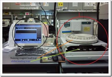
It is a computerized barcode-driven special stain stainer that automatically applies staining reagents to the microscopic slides and mixed over the entire section. Staining process is pre-programmed in the software and accomplished by the serial application of reagents, all of which are held on the reagent carousel (otherwise known as reagent loading tray).

Liquid coverslip is applied before each application of the reagent to inhibit evaporation. Quantity and amount of reagent to be applied on each slide is also pre-programmed by the software.

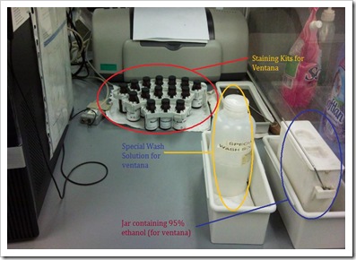
Staining reagent kits are supplied in vials with a barcode labelled carrier, which can be fixed onto the reagent carousel of the machine. The computer ‘recognises’ the barcode and carries out the staining procedure accordingly.

Barcode labels are printed from the software, carrying the biopsy number of the specimen/slide, as well as the barcode specific for performing a particular stain.

Before slides are placed into the machine, it must first be labelled with the barcode labels, and dewaxed. After which, slides are placed into the machine and a special wash solution is added to the slides prior to staining. This aids to prevent sections from drying up.

Appropriate staining kits must be loaded to the reagent carousel, with caps uncapped. Before initialising a run, check that waste bottle is still sufficient for usage, and there is sufficient levels of buffered wash solution and liquid coverslip for the run.

When the machine runs the staining program, it will read off the slide and reagent barcode labels, and information for staining will be downloaded from the system to the staining module to proceed run.

When the run has completed, slides are removed from the machine, and the underside of the slides are gently clean with a gauze to remove residue stain solutions. It is then placed into 95% ethanol to remove excess liquid coverslip, afterwhich, proceeded on to dehydration as per normal. Slides will then be mount with depex, and the machine will be cleaned using a special cleaning kit purchased from the company.

ventana2 ventana1

Pictures taken with permission from lab.

that’s all for now~

Cheers,

Ang Yu Hui

0702632A

Tuesday, September 29, 2009

Reception

Hey everyone! It's my turn (Felicia) to update this blog! ;)

What I'm about to update is on the reception aspect of Haematology.
You may think that the reception does not really play an important role in the entire process of Haematology. In that case, THINK AGAIN!

The reception is one the sub-sections of the Haematology lab and it plays an important role in receiving and processing samples for all the sections in the laboratory. It also handles telephone enquiries from customers for test results. 2 clerks are stationed at the counter. Operating hours for the reception are from 8am to 6pm from Monday to Friday and 8am to 1pm on Saturday.

From 8am to 4pm, each request form received by the counter is clocked-in to initial sample's receiving date and time.

For samples that require the FBC, clerks or technologists manning the counter should check ;

1. That the particulars on the request form tally with the information appearing on the sample label.
2. That the volume of sample is correct and
3. that the tube used is correct.

Once everything is in order, a lab number in ascending order is assigned for FBC samples and written down clearing on the bottom of the left of request form and on the samples tube. The staff who labelled the tube would initial beside the number on the request form. For additional tests, markings are made on the cap of the sample tube so that the technologist is reminded of these tests.

1. O for ESR
2. R for reitculocytes (Retics)
3. MP for blood film malaria (MP)
4. T for HbH Thalassaemia Screening Stage 1

The request forms are logged into the computer using the number assigned. After logging in the request into the computer, the forms together with the samples are brought into the routine lab for processing.

So you see, the reception plays a crutial role in the initial stage of haematological processing! If any data is being logged incorrectly, it may lead to many confusions when being dispatched into the different departments for the required tests.


Felicia
0703345I

Thursday, September 24, 2009

Hi all! Qingling here! I apologise for the late post yeah!


This entry will be on Urine Dipstix!
The urine samples are first collected from the reception by the Health Care Attendant.
Afterwhich, they will tally the name and IC number on the container against the request form. After it is done, he/she will assign a lab number, open up the bottle cap and place them on a tray. 2 rows of 5 bottles each. The request form will be placed under the tray. The med tech will check the request forms to ensure the test ordered is correct. Patients with renal failure or request forms from the Renal Dept has to be tested using the dipsticks. Remove one strip from the dipstick bottle and replace the cap. Completely immerse all the reagent areas of the strip in the urine and remove immediately. While removing it, run the edge of the entire length of the strip against the rim of the urine container to remove excess urine. Then, compare the results with the corresponding color charts on the bottle label.


The intended use of the urine dipstick test is to provide tests for glucose, bilirubin, ketone, specific gravity, blood, pH, protein, urobilinogen, nitrite and leukocytes in urine.
The tips of the dipstick are impregnated with chemicals which react with abnormal substances in the urine to produce coloured end products. In some of the tests, the depth of the color produced is related to the concentration of the abnormal substances in urine.


Results should be reported in this manner :
For pH, the glomerulur filtrate of blood plasma is usually acidified by renal tubules and collecting ducts from a pH of 7.4 to about 6.0 in the final urine. Both normal and abnormal range is from 5 to 9.

For specific gravity, it measures the urine density. Random urine may vary from 1.001 - 1.035. Normal adults' urine with normal diet and fluid intake will have SG of 1.016 - 1.3022. Urine pH above 1.035 is either contaminated or have high glucose level.

For protein, normal protein excretion does not usually exceed 150mg/24hours or 10mg/100mL in any single specimen. Normally, no protein is detectable in urine. Color matching greater than Trace indicates proteinuria.

For glucose, dipsticks employing the glucose oxidation reaction for screening are specific for glucose but can miss other reducing sugars such as galactose and fructose.

For ketones, it should be negative in normal urine specimens. Detectable level of ketone may occur in urine during physiological stress such as fasting, pregnancy and vigorous exercise. In ketoacidosis, starvation or with other abnormalities of carbohydrate or lipid metabolism, ketones may appear in large amount before serum ketone concentration are elevated.

For blood, the significance may vary among patients. Blood is often but not always found in urine of menstruating females. It is highly sensitive to haemoglobin and thus complements the microscopic examination.

For bilirubin, there should be no bilirubin detected in normal individual. Trace amount are sufficiently abnormal to require further investigations. Thus colours that are unlike the negative or positive colour may indicate that bilirubin-derived bile pigments are present in urine sample and may be masking the bilirubin reaction.

For urobilinogen, it will detect the urobilinogen in concentration as low as 3umol/L in urine. A result of 33umol/L represents the transition from normal to abnormal.

For nitrite, it should not be detected in normal individual. Proportion of positive nitrite tests in cases of significant infection depends on how long the urine specimens were retained in the bladder prior collection.

For leukocytes, normal urine usually have negative result. Positive results are clinically significant and may be found in random specimens from female due to contamination of specimen by vaginal discharge.

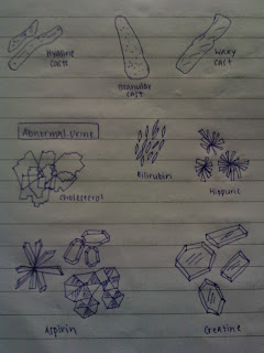
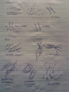
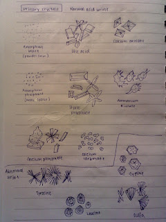
Here is what i've drawn in my own notebook. This are pictures of how cells look like under microscope! Its not very bright. Sorry!


Thats basically all! Feel free to ask if you have any doubts! :D


Sunday, September 6, 2009

MTS assay

CellTiter 96® AQueous One Solution Cell Proliferation assay (MTS assay)
It is a colorimetric method for determining the number of viable cells in proliferation or cytotoxicity assays.

MTS tetrazolium compound i.e. (3-(4,5-dimethylthiazol-2-yl)-5-(3-carboxymethoxyphenyl)-2-(4-sulfophenyl)-2H-tetrazolium) is bioreduced by cells intro a coloured formazan product, which is soluble in tissue culture medium – decreasing toxicity to cells seen with an insoluble product (MTT assay). This conversion is accomplished by NADPH or NADH produced by dehydrogenase enzymes in metabolically active cells
Phenazine methosulfate (PMS), an electron-coupling reagent, is combined with MTS reagent to form a stable solution due to PMS’s enhanced stability.
The quantitiy of formazan product is directly proportional to the number of viable cells in the culture as measured by the absorbance at 490nm.

For the MTS assay, 5 x 10³ cells per well were cultured in 96-well plates and treated with cytotoxic compounds (What I am studying) for 48 and 72 hours respectively. After incubation for specified times at 37°C in a humidified incubator, 25µL of MTS reagent was added to each well and further incubated for 2-4 hours. Absorbance was measured at 490 nm on a micro plate reader.

Advantages
• Fast – Eliminates solubilization of formazan crystals before absorbance reading and MTS is more efficiently bioreduced compared to MTT
• Safe – Requires no volatile (easily evaporated at normal temperatures) solvent to solubilize formazan product

Li Yinliang Alex 0704894E
TG02 Group 8
6 September 2009